Rapp OptoElectronic
  • Home
  • Products
    • Photomanipulation & Microdissection (LCM)
      • Scanner-Based Photomanipulation – UGA-42 Series
        • UGA-42 Firefly
        • UGA-42 Geo
        • UGA-42 Caliburn
      • Fixed-Spot Photomanipulation – AiWon Series
        • AiWon
        • AiWon Geo
        • AiWon Caliburn
      • Advanced Photomanipulation
        • FLUCS Micro Flow Photomanipulation
        • HOLO4D Holographic Photomanipulation
      • Laser Capture Microdissection (LCM) Accuva Cellect
    • Microscopy & Imaging
      • Multi-Photon Microscopy
        • MOM
        • DF-Scope
      • Raman Microscopy
        • XperRAM C Series
      • Deep UV Microscopy
      • Spinning Disk Microscopy
        • X-Light V2
      • Further Microscopy Systems
        • BOB
        • SOM
        • OpenFrame
    • Microscope Accessories
      • Optical Couplings
        • IX3-Breadboard
        • FC-70 Series
        • LGA
      • Camera Adapter
        • IX3-CAM Adapter
        • TwinCam
        • MultiCam
      • Image Splitter
        • OptoSplit II
        • OptoSplit II BP
        • OptoSplit III
        • MultiSplit V2
    • Illumination Systems
      • LED & Bulb Systems
        • KSL2
        • UVILED
        • UVICO-2
        • FuraLED
        • OptoScan
      • Laser Systems
        • Single Line Laser
        • Laser Combiner
      • Flash Lamp Systems
        • JML-C2
        • SP-20
  • Applications
    • Ablation & Microdissection
    • DNA Damage
    • IR Temperature Jump
    • Uncaging & Photolysis
    • Optogenetics & Photostimulation
    • Photobleaching & FRAP
    • Photoconversion & Photoswitching
  • Publications
    • Ablation & Microdissection
    • DNA Damage
    • IR Temperature Jump
    • Uncaging & Photolysis
    • Optogenetics & Photostimulation
    • Photobleaching & FRAP
    • Multi-Photon Imaging
    • Microscope Illumination
    • FLUCS Micro Flow
  • About us
    • Partners
    • Team
    • Our story
    • Location
  • Events
  • Contact
  • Career
  • Click to open the search input field Click to open the search input field Search
  • Menu Menu

Investigation of Protein Dynamics in Focal Adhesions Using FRAP and TIRF

Photobleaching & FRAP

Mouse embryonic fibroblasts (MEF) were transfected with different mutations of the adaptor protein vinculin and FRAP experiments were performed to analyze the influence of the mutations on the incorporation of vinculin into focal adhesions (FAs).

For the quantification of the real protein exchange dynamics within focal adhesions without overlying cytosolic diffusion artifacts, the FRAP technique was combined with TIRF (total internal reflection fluorescence) microscopy. To this end, a Zeiss Observer Z.1 (TIRF) microscope was equipped with a Rapp OptoElectronic fixed spot illumination device coupled to a 473 nm diode laser (DL-473). In combination with a 100x objective, a spot size of approximately 5 µm in diameter was illuminated, allowing bleaching of single focal adhesions in living cells. The results indicate a slight, but significant increase in mobile fraction for the Y1065E-mutant and an uncoordinated incorporation of the Y1065F-mutant into FAs.

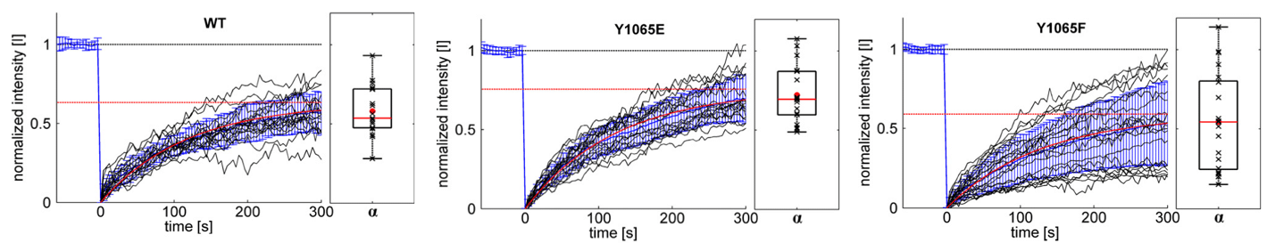
Figure 1: Exchange dynamics of different vinculin constructs measured with a TIRF-FRAP setup. Different eGFP-vinculin constructs (WT, Y1065E and Y1065F) were expressed in vin‑/‑ MEFs. Before bleaching, cells were monitored for at least 5 minutes to ensure that only stable FAs were analyzed. Black graphs show the normalized intensity [I] curves from individual measurements. The mean value and its standard deviation σ are shown in blue; results from the fit of mean values to the kinetic model are shown in red. The saturation value α indicates the mobile fraction (red dotted line). At the right of each graph, the box plots show the distribution of the mobile fractions of single measurements (black “x”). The mean values are given as a red dot, the median as a red line.


Setup:

  • Microscope: Zeiss Observer Z.1 with TIRF
  • Objective: 100x α-Plan-Apochromat (oil) NA 1.46

Rapp OptoElectronic components:

  • FRAP System: ZSI fixed spot illumination
  • Light source: DL-473 diode laser
  • Optical fiber: multimode (ø = 550 µm) => spot size on sample approx. 5 µm


Published:

Küpper at al. 2010. “Tyrosine Phosphorylation of Vinculin at Position 1065 Modifies Focal Adhesion Dynamics and Cell Tractions.” Biochemical and Biophysical Research Communications 399 (4): 560–64. doi:10.1016/j.bbrc.2010.07.110.

https://rapp-opto.com/wp-content/uploads/2018/10/kuepper-frap-icon.png 204 354 Anette https://rapp-opto.com/wp-content/uploads/2023/05/rapp-logo-340pxblau-300x79.png Anette2018-10-22 14:18:112019-03-27 11:53:20Investigation of Protein Dynamics in Focal Adhesions Using FRAP and TIRF

Latest Applications

  • FLIRT: fast local infrared thermogenetics for subcellular control of protein function8. June 2021 - 15:31
  • Following Tubulin Dynamics during Spindle Formation using Photoconversion of mEos223. October 2018 - 18:25
  • Optogenetical Stimulation in Acute Brain Slices23. October 2018 - 15:43

Applications Categories

  • Ablation & Microdissection
  • DNA-Damage
  • Temperature Jump & IR-Lego
  • Uncaging & Photolysis
  • Optogenetics & Photostimulation
  • Photobleaching & FRAP
  • Photoconversion &-switching
Copyright © 2026
  • Contact
  • Newsletter
  • Imprint
  • Privacy Policy
  • Sitemap
Scroll to top Scroll to top Scroll to top