SOM®
Simple Moving Microscope
Sutter Instrument
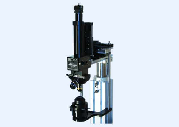
The Son of MOM® (SOM®) is a small, simple microscope. It allows to use a single experimental setup for both: in vivo and in vitro experimentation. As in the two-photon Movable Objective Microscope (MOM), positioning over the sample and focusing is accomplished automatically. This removes the need for large translators and stages that normally limit the available space beneath the objective for in vivo experimentation. For example, the SOM would allow for whole-cell patch recordings from neurons in vivo on one day followed by multi-cell recordings in slices on the next.
SOM opens up experimental possibilities that otherwise might be limited by the ever growing space constraints in modern laboratories. The SOM is designed to take full advantage of our free Multi-Link™ software program for micromanipulator positioning.
For instance, during whole-cell patch recordings in slices, it is commonly necessary to search within a large tissue area to find neurons appropriate to your experiment. With SOM, you simply translate over your sample to search for your target. The software programs will then retrieve your recording and stimulation pipettes, allowing you to start recording immediately. Moreover, if you need to stimulate a region outside of the current objective’s field of view, the programs will allow you to lock the position of your recording pipette and reposition the objective and stimulating pipette(s) to their required positions.
An optional Oblique Coherent Contrast (OCC) condenser that is illuminated with an LED is also available. The condenser translates with the microscope in the X & Y axes, which allows for consistent illumination during re-positioning of the SOM over the sample.
How it Works:
The SOM is designed to take advantage of the high-quality images that can be obtained with a simple IR LED-based transmitted light source combined with an IR capable CCD camera. This combination is sufficient for the majority of in vitro electrophysiology needs. The SOM is also designed with a two-position filter cube allowing identification of fluorescently-tagged cells for recording or for photostimulation. If you equip both of the filter cube positions, one of the filter sets will need to pass IR to allow for transmitted light imaging. As many filter combinations will pass IR, transmitted light imaging can generally be done in either of the two filter positions.
The fluorescence excitation port of the microscope has C-mount threading as well as mounting holes for standard cage components. This allows for customization by the user to various experimental needs. For instance, multiple light sources can be coupled to the excitation port with small cage assemblies.
- Simple moving microscope based on an MP-285/MPC-385 motorized micromanipulator
- X, Y and Z axes of manipulator used to position microscope over the sample and focus. No need for large translators or moving stages
- Optimized to allow in vivo and in vitro experimentation on one setup
- Standard configuration accepts RMS thread objectives – Contact us for additional options
- Free Multi-Link™ software coordinates movement with micropipette positioning of MPC-200
- Transmitted IR- and epi-fluorescent imaging modes
- Flexible excitation port allows easy addition of secondary sources for photostimulation
- MPC-200 controller with USB interface and open source commands


