Rapp OptoElectronic
  • Home
  • Products
    • Photomanipulation
      • Scanner-Based Photomanipulation – UGA-42 Series
        • UGA-42 Firefly
        • UGA-42 Geo
        • UGA-42 Caliburn
      • Fixed-Spot Photomanipulation – AiWon Series
        • AiWon
        • AiWon Geo
        • AiWon Caliburn
      • FLUCS Micro flow photomanipulation
    • Microscopy & Imaging
      • Multi-Photon Microscopy
        • MOM
        • DF-Scope
      • Spinning Disk Microscopy
        • X-Light V2
      • Further Microscopy Systems
        • BOB
        • SOM
      • Microscope Modification
    • Microscope Accessories
      • Optical Couplings
        • IX3-Breadboard
        • FC-70 Series
        • LGA
      • Camera Adapter
        • IX3-CAM Adapter
        • TwinCam
        • MultiCam
      • Image Splitter
        • OptoSplit II
        • OptoSplit II BP
        • OptoSplit III
        • MultiSplit V2
    • Illumination Systems
      • LED & Bulb Systems
        • KSL2
        • UVILED
        • UVICO-2
        • FuraLED
        • OptoScan
      • Laser Systems
        • Single Line Laser
        • Laser Combiner
      • Flash Lamp Systems
        • JML-C2
        • SP-20
  • Applications
    • Ablation & Microdissection
    • DNA Damage
    • IR Temperature Jump
    • Uncaging & Photolysis
    • Optogenetics & Photostimulation
    • Photobleaching & FRAP
    • Photoconversion & Photoswitching
  • Publications
    • Ablation & Microdissection
    • DNA Damage
    • IR Temperature Jump
    • Uncaging & Photolysis
    • Optogenetics & Photostimulation
    • Photobleaching & FRAP
    • Multi-Photon Imaging
    • Microscope Illumination
  • About us
    • Partners
    • Team
    • Our story
    • Location
  • Events
  • Contact
  • Career
  • Search
  • Menu Menu

Multi-Photon Microscopy

Photon-Based Deep Tissue Imaging

Multi-photon microscopy enables an increased penetration depth into the sample tissue by stimulating fluorescence in the near IR window of biological tissue, and by providing a distinct advantage in three-dimensional imaging. The low absorption of the stimulation light in the tissue avoids photobleaching and phototoxicity, while the multi-photon effect leads to a sharper definition of the focal plane, reducing background fluorescence. Multi-photon microscopy is the method of choice for three-dimensional imaging of living cell structures, such as brain slices, or in vivo embryos and adult animals.

The following multiphoton systems are available:

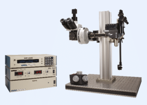
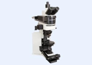
  • MOM® – Movable Objective Microscope system
  • DF-ScopeTM – Multi-photon imaging package for upright microscopes
Link to: MOM

MOM

Link to: DF-Scope

DF-Scope

Applications

  • In vivo multi-photon imaging
  • Electrophysiological recording and imaging (culture, large in vivo preparations, etc.)
  • Non-horizontal surface microscopy
  • Simultaneous retinal stimulation and two-photon microscopy
  • Whole animal imaging
  • Immunology
  • Embryology

Copyright © 2023
  • Contact
  • Newsletter
  • Imprint
  • Privacy Policy
  • Sitemap
X
Scroll to top