Rapp OptoElectronic
  • Home
  • Products
    • Photomanipulation & Microdissection (LCM)
      • Scanner-Based Photomanipulation – UGA-42 Series
        • UGA-42 Firefly
        • UGA-42 Geo
        • UGA-42 Caliburn
      • Fixed-Spot Photomanipulation – AiWon Series
        • AiWon
        • AiWon Geo
        • AiWon Caliburn
      • Advanced Photomanipulation
        • FLUCS Micro Flow Photomanipulation
        • HOLO4D Holographic Photomanipulation
      • Laser Capture Microdissection (LCM) Accuva Cellect
    • Microscopy & Imaging
      • Multi-Photon Microscopy
        • MOM
        • DF-Scope
      • Raman Microscopy
        • XperRAM C Series
      • Deep UV Microscopy
      • Spinning Disk Microscopy
        • X-Light V2
      • Further Microscopy Systems
        • BOB
        • SOM
        • OpenFrame
    • Microscope Accessories
      • Optical Couplings
        • IX3-Breadboard
        • FC-70 Series
        • LGA
      • Camera Adapter
        • IX3-CAM Adapter
        • TwinCam
        • MultiCam
      • Image Splitter
        • OptoSplit II
        • OptoSplit II BP
        • OptoSplit III
        • MultiSplit V2
    • Illumination Systems
      • LED & Bulb Systems
        • KSL2
        • UVILED
        • UVICO-2
        • FuraLED
        • OptoScan
      • Laser Systems
        • Single Line Laser
        • Laser Combiner
      • Flash Lamp Systems
        • JML-C2
        • SP-20
  • Applications
    • Ablation & Microdissection
    • DNA Damage
    • IR Temperature Jump
    • Uncaging & Photolysis
    • Optogenetics & Photostimulation
    • Photobleaching & FRAP
    • Photoconversion & Photoswitching
  • Publications
    • Ablation & Microdissection
    • DNA Damage
    • IR Temperature Jump
    • Uncaging & Photolysis
    • Optogenetics & Photostimulation
    • Photobleaching & FRAP
    • Multi-Photon Imaging
    • Microscope Illumination
    • FLUCS Micro Flow
  • About us
    • Partners
    • Team
    • Our story
    • Location
  • Events
  • Contact
  • Career
  • Search
  • Menu Menu

OptoSplit II

Dual Emission Image Splitter
Cairn Research Ltd.

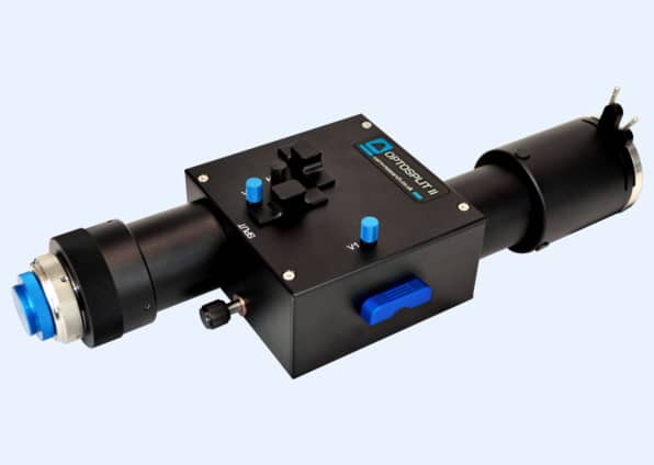
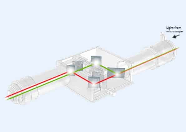
The dual emission image splitter OptoSplit II, a product of our partner company Cairn Research Ltd., is a simple device enabling a single camera to record images simultaneously at two different optical wavelengths, polarization states or other differentiated states. To improve the usability for multi-user microscopes, please have a look at the OptoSplit II with bypass option (OptoSplit II BP).

Features

Fast and Low Cost Simultaneous Imaging

Traditionally, dual channel imaging is performed using an electronic filter changer or an additional camera and beam splitter, neither of which is ideal for all applications. The switching speed of an electronic filter changer limits the temporal resolution, whereas a second camera adds cost and complexity. In comparison, the OptoSplit II is a low cost option for simultaneous imaging without any time delays between the acquired channels.

Easy and Optimal Alignment

The OptoSplit II uses a unique rotating mirror cradle, which gives adjustable spatial separation to ensure excellent image registration. It features a fully adjustable rectangular aperture to enable cropped sensor imaging modes and reduced scatter. The adjustment is quite simple and allows to setup the module on your microscopic system by yourself.

For Large Camera Sensors

The latest version uses our own lens design to support larger camera sensors of scientific CMOS and EMCCD cameras. The instruments have a correspondingly larger aperture and improved off-axis correction to give enhanced performance with all camera sensors.

Applications
  • Simultaneous multi fluorescent probe imaging
  • Simultaneous phase contrast/ DIC and fluorescence
  • Simultaneous multi depth imaging (using independent lenses)
  • Förster Resonance Energy Transfer (FRET)
  • Ratiometric calcium, voltage and pH-imaging (https://www.youtube.com/watch?v=GKNFYARHfcQ)
  • Photoconversion/ Photoswitching
  • Total Internal Reflection Fluorescence (TIRF)
  • Spinning disk confocal microscopy
  • Single Plane Illumination Microscopy (SPIM)
  • Polarization studies (Anisotropy)
  • 3D super resolution PALM/ STORM (using cylindrical lenses)
Specifications
  • Compact design with integral C-mount input and output ports
  • Simple and precise controls for image registration
  • Interchangeable filter/ dichroic holders
  • Rotating filter mount for polarization studies
  • 1x, 1.3x and 1.7x magnification available
  • Support for sensors up to 18.8 mm diagonal (13.3 mm x 13.3 mm sensors)
  • Angled and flat auxiliary drop ins for neutral density filtering, polarizers or chromatic correction
  • Supports 25 mm filters
  • Supports 2 mm thick Ultra-flat dichroics along with our recommended size (26x38x2 mm)
  • 40 mm diameter proprietary optics
  • 425 nm to 875 nm coatings on all surfaces
Downloads
  • OptoSplit II Flyer (579.2 KB)
  • OptoSplit II Manual (1.1 MB)
  • Support video (tutorial):
https://www.youtube.com/watch?v=T_TtQHQpAkA
Literature

Zanetti-Domingues LC, Korovesis D, Needham SR, Tynan CJ, Sagawa S, Roberts SK, Kuzmanic A, Ortiz-Zapater E, Jain P, Roovers RC, Lajevardipour A, van Bergen En Henegouwen PMP, Santis G, Clayton AHA, Clarke DT, Gervasio FL, Shan Y, Shaw DE, Rolfe DJ, Parker PJ and Martin-Fernandez ML. (2018) The architecture of EGFR’s basal complexes reveals autoinhibition mechanisms in dimers and oligomers. Nat Commun. Oct 18; 9(1):4325.

Hu J, Wu M, Jiang L, Zhong Z, Zhou Z, Rujiralai T and Ma J. (2018) Combining gold nanoparticle antennas with single-molecule fluorescence resonance energy transfer (smFRET) to study DNA hairpin dynamics. Nanoscale. Apr 5;10(14):6611-6619.

Cavadini P, Weinhold H, Tönsmann M, Chilingaryan S, Kopmann A, Lewkowicz A, Miao C, Scharfer P and Schabel W. (2018) Investigation of the flow structure in thin polymer films using 3D µPTV enhanced by GPU. Experiments in Fluids 59:61(4)

Bizebard T, Arluison V and Bockelmann U. (2018) Single-Molecule FRET Assay to Observe the Activity of Proteins Involved in RNA/RNA Annealing. Methods Mol Biol.; 1737:301-319.

Wasim L and Treanor B. (2018) Single-Particle Tracking of Cell Surface Proteins. B Cell Receptor Signaling pp 183-192, Feb.; part of the Methods in Molecular Biology book series (MIMB, volume 1707)

He H, Ye Z, Xiao Y, Yang W, Qian X and Yang Y. (2018) Super-Resolution Monitoring of Mitochondrial Dynamics upon Time-Gated Photo-Triggered Release of Nitric Oxide. Anal Chem. Feb 6; 90(3):2164-2169.

Schrangl L, Göhring J and Schütz GJ. (2018) Kinetic analysis of single molecule FRET transitions without trajectories. J Chem Phys. Mar 28; 148(12):123328.

Wang S, Brettmann JB and Nichols CG. (2018) Studying Structural Dynamics of Potassium Channels by Single-Molecule FRET. Methods Mol Biol.1684:163-180.

Van Krugten J and Peterman EJG. (2018) Single-Molecule Fluorescence Microscopy in Living Caenorhabditis elegans. Methods Mol Biol. 1665:145-154.

Zhao YT, Guo YB, Fan XX, Yang HQ, Zhou P, Chen Z, Yuan Q, Ye H, Ji GJand Wang SQ. (2017) Role of FK506-binding protein in Ca2+ Spark regulation. Science Bulletin 62:19; 1295-1303.

Copyright © 2025
  • Contact
  • Newsletter
  • Imprint
  • Privacy Policy
  • Sitemap
Scroll to top