Rapp OptoElectronic
  • Home
  • Products
    • Photomanipulation
      • Scanner-Based Photomanipulation – UGA-42 Series
        • UGA-42 Firefly
        • UGA-42 Geo
        • UGA-42 Caliburn
      • Fixed-Spot Photomanipulation – AiWon Series
        • AiWon
        • AiWon Geo
        • AiWon Caliburn
      • FLUCS Micro flow photomanipulation
    • Microscopy & Imaging
      • Multi-Photon Microscopy
        • MOM
        • DF-Scope
      • Spinning Disk Microscopy
        • X-Light V2
      • Further Microscopy Systems
        • BOB
        • SOM
      • Microscope Modification
    • Microscope Accessories
      • Optical Couplings
        • IX3-Breadboard
        • FC-70 Series
        • LGA
      • Camera Adapter
        • IX3-CAM Adapter
        • TwinCam
        • MultiCam
      • Image Splitter
        • OptoSplit II
        • OptoSplit II BP
        • OptoSplit III
        • MultiSplit V2
    • Illumination Systems
      • LED & Bulb Systems
        • KSL2
        • UVILED
        • UVICO-2
        • FuraLED
        • OptoScan
      • Laser Systems
        • Single Line Laser
        • Laser Combiner
      • Flash Lamp Systems
        • JML-C2
        • SP-20
  • Applications
    • Ablation & Microdissection
    • DNA Damage
    • IR Temperature Jump
    • Uncaging & Photolysis
    • Optogenetics & Photostimulation
    • Photobleaching & FRAP
    • Photoconversion & Photoswitching
  • Publications
    • Ablation & Microdissection
    • DNA Damage
    • IR Temperature Jump
    • Uncaging & Photolysis
    • Optogenetics & Photostimulation
    • Photobleaching & FRAP
    • Multi-Photon Imaging
    • Microscope Illumination
  • About us
    • Partners
    • Team
    • Our story
    • Location
  • Events
  • Contact
  • Career
  • Search
  • Menu Menu

MOM

Movable Objective Microscope
Sutter Instrument

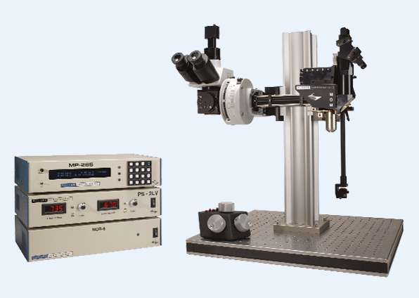
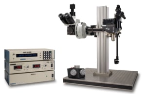
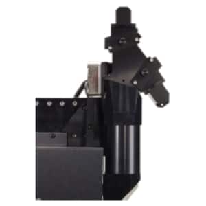
The Movable Objective Microscope® (MOM®) is a two-photon microscope capable of imaging deep within living specimens when combined with a Ti:Sapphire Laser. The MOM design is unique in providing three-dimensional objective movement and rotation allowing the specimen to remain stationary. Many highly regarded imaging laboratories around the world use the Sutter MOM and we constantly work with our customers to adapt the design for their changing needs.

Opto-mechanical Design

Opto-mechanical Design

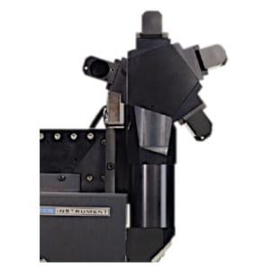
The MOM consists of two independent microscopes. The wide-field half of the microscope consists of an Olympus vertical illuminator, Sutter Xenon arc lamp and camera mount to provide standard epifluorescence. The two-photon side of the microscope provides the optical pathway for guiding the excitation laser light from the table up into the scanning galvanometric mirrors. From there the beam expands through the scan lens and directs into the back of the objective. Following two-photon excitation, the emitted photons are directed into the detection pathway by a dichroic mirror immediately above the objective. The main body of the microscope moves backwards on a rail system allowing easy access to the specimen prior to imaging.

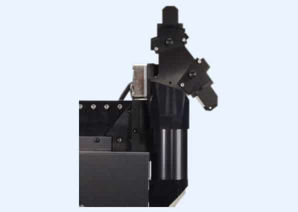
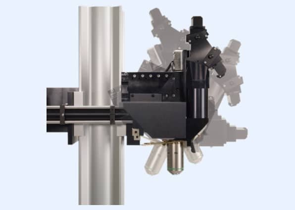
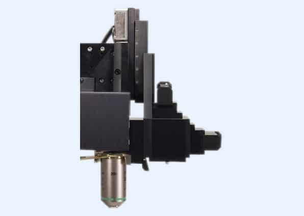
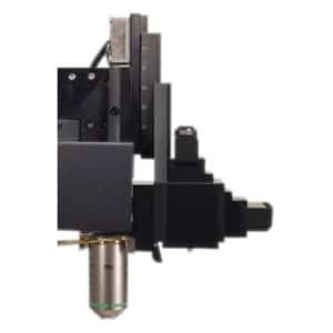
The objective translates in X, Y and Z axes, it additionally rotates around the X axis. Two moving mirrors allow the microscope to maintain efficient delivery of the excitation light to the back aperture of the objective regardless of movement or orientation. The X, Y and Z movements used are the same as controlled in our MP-285 micromanipulator, so you know the movements are smooth, fine in scale, drift-free and highly reproducible. These movements allow the acquisition of Z-stacks and mosaic images of large regions of tissue without the need for a moving stage.

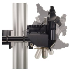
The horizontal light path allows for rotation of the objective away from the standard vertical position. As a result of this rotation, the MOM can easily be converted from an upright to an inverted microscope and the objective positioned from 0 to 180 degrees. This positional freedom permits the imaging of non-horizontal surfaces and volumes.

Scanning Systems

Scanning Systems

During the last 10 years, scanning technology has changed dramatically: objectives changed requiring larger beam sizes and scanner technology advanced providing dependable resonant scanners. Unlike other two-photon microscope designs, the MOM has lived through and adapted to changes in scanning technology. Throughout this development, Sutter has maintained two principles. First, existing scopes can be upgraded to new technology as it becomes available. Many original scopes with 3 mm galvo scanners have been upgraded to either 6 mm galvo scanners or resonant/ galvo scanners. Second, Sutter continues to supply the original designs if they are needed for current research. We can provide a 3 mm or 6 mm conventional scanning MOM or a resonant/ galvo scanning MOM all at competitive prices.

The MOM is available with four different detector path designs. The original two channel pentagon, which can become a four channel detector path. The Short path and the Wide path are two designs that increase the chances of capturing ballistic photons by getting the first collection lens either closer to the back aperture of the objective (Short path) or by using a larger aperture collection lens and dichroic (Wide path).

Sutter MOM packages include all of the equipment (less the Ti:Sapphire laser and objective) needed for a complete imaging system.

  • Cambridge Technology XY galvanometric and resonant scanners
    (conventional with 3 or 6 mm mirrors or resonant with 5mm mirrors)
  • Hamamatsu photomultiplier tubes (PMTs):
    R6357 multialkali or H10770PA-40 (GaAsP) products
    (Sutter is an authorized reseller for Hamamatsu)
  • Power supplies for PMTs:
    Either a Sutter PS-2 (dual channel high-voltage power supply for R6357 PMTs) or
    Sutter PS-2LV (dual channel low-voltage power supply for H10770PA-40 (GaAsP) PMTs) can be ordered
  • Hamamatsu, Sigmann or FEMTO pre-amplifiers, selection varies with software and type of scan
  • Data acquisition: National Instruments and Measurement Computing systems
Features
  • Objective moves 22 mm in X, Y and Z
  • Objective rotates about optical axis for imaging of non-horizontal surfaces and volumes
  • Customizable open platform design
  • Cambridge Technology conventional or resonant XY scanners
  • Two or four channel detector system with Hamamatsu PMTs and preamplifiers
  • Sutter PS-2/ PS-2LV dual channel PMT power supply
  • National Instruments/ Measurement Computing based data acquisition systems
Applications
  • In vivo two-photon imaging
  • Electrophysiological recording and imaging (culture, large in vivo preparations, etc.)
  • Non-horizontal surface microscopy
  • Simultaneous retinal stimulation and two-photon microscopy1
  • Whole animal imaging
  • Immunology
  • Embryology

(1) “Eyecup scope-optical recordings of light stimulus-evoked fluorescence signals in the retina”, Euler et al., Pflugers Arch, 2008

Software

Starting in 2011, Sutter began offering the MOM Computer System and Software (MCS). Before this software package was developed, most users relied on ScanImage or MPScope to generate scanned images. Customers valued the fact that the MOM would operate with open source freewares, however, there seemed to also be a market for a commercial package. Sutter MScan offered a number of functions that were not available in the existing freeware packages including photostimulation and the ability to combine imaging with electrophysiological recording and photostimulation. As resonant scanning became popular, and none of the freewares supported resonant scanning on the MOM, Sutter and MScan took the initiative. The latest version, MScan 2.0, coupled with a faster data acquisition system, allowed the MOM to generate fast, resonant images. To this day, the Sutter MOM with MScan2.0 remains the one platform available that can switch back and forth between conventional and resonant scanning.

The MOM® has always been compatible with ScanImage freeware, the two-photon imaging software developed by Karel Svoboda and collaborators. One of the reasons the MOM platform exists in its present form is the strong support from the ScanImage community. In 2014, Vidrio became the principle vehicle for support and new development of ScanImage. Sutter is happy to make Vidrio ScanImage Premium available to customers who wish premium support and the latest features. ScanImage freeware is also still available as SI5. Sutter provides packages that include the necessary data acquisition hardware to couple the MOM and other scanning microscopes to ScanImage Premium or SI5.

Downloads
  • MOM® Flyer
  • ScanImage Flyer

Copyright © 2023
  • Contact
  • Newsletter
  • Imprint
  • Privacy Policy
  • Sitemap
X
Scroll to top